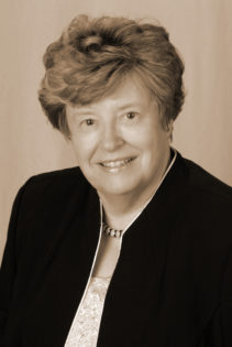
 It was truly an honor to interview Joan P. Baker MSR, RDMS, RDCS, FSDMS. Originally from England, Baker was invited to the United States in the 1960s – due to her sonography passion and practice – and she’s been here ever since.
Baker’s eclectic career as a clinical sonographer, educator, and now president of the company Sound Ergonomics has many highlights:
- She helped create the Society of Diagnostic Medical Sonography (SDMS) and the American Registry for Diagnostic Medical Sonographers (ARDMS)
- She testified in front of the U.S. Office of Education so that sonography could be officially recognized as a profession
- Baker was named a sonography pioneer by the Smithsonian Institute, acting as a legal expert in sonography cases
- She testified in front of the federal Occupational Safety and Health Administration
It is no surprise that the SDMS has created an award in her name (the Joan P. Baker Pioneer Award.)!
In this interview, Baker talks about how she in fact pioneered the profession, how ultrasound has evolved over time, and the importance for Sonographers to scan safely.
JB: Well, it really wasn’t a decision – it was by default. I was the last hired and the least qualified, and the dubious honor for that is you get to do something that other people have tried and considered to be a waste of time. So I was the last hired and the least qualified at the National Hospital for Nervous Diseases [now called the National Hospital for Neurology and Neuroscience] in Queens Square, London, which is a famous institution. I ended up being the one that got to do nuclear medicine and ultrasound.
JB: Yes or they didn’t even give them a look. This was 1960 so [ultrasound] was not on the market. It was a prototype and it didn’t look very attractive. There were no sonographers at the time. There was nobody except a handful of physicians in the world and a handful of engineers who were working with those physicians. And typically you didn’t know about them because there wasn’t the communication mechanisms that there are today…I knew about Professor Ian Donald, for example, who was in Glasgow Scotland at the time – we’ later became acquainted – but at that time he too was a lone wolf using ultrasound in both obstetrics and gynecology. He was performing ultrasound using B-scan. I was using A-mode only. All I saw on my screen were spikes.
JB: Well I’d been [at the National Hospital] for four years when I got an invitation from a Dr. Leslie Zatz, a neuroradiologist at the Stanford Medical Center in Palo Alto, California, asking me if I’d like to come over to America to Stanford and do ultrasound for them. So I looked up this place called ‘Stanford Medical Center’ – I was 23 years of age – and I found it was quite a prestigious institution. I assumed I was going to join the best place to do ultrasound in the world. And when I got there, Dr Zatz grinned at me and proudly told me he found the on/off switch…I was specifically asked to go for a year to Stanford Medical Center. I was young and naïve and at the same time excited and honored to be asked.
JB: Well it was I suppose, but when you’re only 23 years old, you have a different perspective. You’re invincible and you don’t look at things from the negative side. You look at things from the positive side. I had an identical twin and two brothers, and a mother and father that I was leaving, and knew nobody in the United States…I never gave a thought to the fact that my parents might be affected by this. I was just so excited to go that I thought they would be equally excited for me. For all intents and purposes that’s what they showed, but they knew that if I stayed much longer than a year that I would probably not come home. Certainly you can say I did overstay my welcome. It was arranged for a year, and I stayed for a year-and-a-half, and then I didn’t go back [to England except] to visit.
JB: [laughs] From 1960 to 2018, yes. From A-mode, to B-mode, to real-time, to gray scale, oh yes, just enormous. The first ultrasound machine I used to scan at Stanford… actually filled a 10 by 10-foot room. Obviously there was no portability at all. But then the ultrasound machine I worked with in 1960 in London had portability because it was on a cart-based system on wheels, but it was just an A-mode.
JB: Yes, unless you consider teaching a different field. I mean I taught ultrasound and nuclear medicine. I have educated somewhere between 400 and 600 students. I was contacted by some of my graduates who told me they were scanning in pain. This shocked me, so for the latter part of my career I have studied and been devoted to reducing the incidence of occupational injury in sonography.
JB: Yes it did. One thing we do is go to [diagnostic imaging workplaces] and we’ll do assessments. We will take pictures of sonographers scanning and advise them on how they could improve their technique to hopefully lesson the pain and suffering that they’re going through…Now 90 percent of sonographers are scanning in pain or discomfort.
As far as prevention is concerned, we do put out information for students in ultrasound and for [educational] programs…so that they can perhaps prevent injuries – that’s the best thing to do is prevent it from happening…A lot of [educational] program directors and faculty are in that position because their didactic career ended because of injury. So that makes some of them very devoted to teaching it and making sure their students don’t end up the way they do. But on the other hand, some of them say “that comes with the job,” which is very sad to hear.
What we have found is it takes the commitment of administrators manufacturers and sonographers to mitigate workplace injury and to actually make a change and make a difference. And if one of the legs of that stool isn’t in place then nothing positive is going to come out of it.
We work also with the OEMS (commercial companies) to help them design the control panel and the equipment, to make it more ergonomic, more user-friendly.
Sadly administration buys the equipment that’s got the cheapest price tag on it. Invariably that is one that has cut the corners intentionally. [The manufacturer] probably offers a very ergonomic piece of equipment but that’s not the one the customer typically buys. The customer buys the midrange equipment that doesn’t have the bells and whistles on it, and then they expect the sonographer to use it as if it were an ergonomic piece that costs twice as much.
JB: A lot of sonographers say to us, “No it’s not my right side that I scan with that hurts, it’s my left side.” If your right side hurts, you lean over more to the right in order to compensate for the pain in your right shoulder. When you do that now you overstretch with your left arm in order to reach the control panel. We call that position The Crucifixion.
JB: You can’t [scan] ergonomically every time. But the point is if you don’t know what is the right way ergonomically you’ll unlikely ever do it the right way. If you know what is right and you know what to avoid, then you do your very best to do it as much as you can in that way. And this will lessen your chances of injury.
Then if you get the scheduling done right so that you don’t schedule back-to-back difficult cases, like multiple gestations back-to-back… There are lots of “multiple” things that are very much more injury-producing, but the scheduler doesn’t consider that, they just schedule them.
There’s two ways to run a lab. If you run a self-paced lab that is a lab where the sonographer takes the patient in and does the scan, and takes their time and does a good job, and comes out and does something else, like putting the case together and writing a preliminary report in an ergonomic way…and then they go and get the next patient (their muscles ad tendons have had a chance to recover). But if you’ve got a lab that gives a list of patient names to the sonographer when they walk into work in the morning and they say, “That’s your day’s work”…and it could be that they do a whole day of portables. So for a whole day, non-stop they’re going from room to room up on the floor doing patients in their beds, which is very difficult to do ergonomically…or they get add-ons that increase their patient load.
JB: I think creating the occupation of diagnostic medical sonographer would be a highlight and a victory…It put ten years on my life…It was in front of the United States Office of Education, and the stress levels are running high because you’re there trying to do your best for a lot of people…who are counting on you to succeed.
…There were about 23 allied health professions at the time and there were two or three new ones going up for the first time. And we were doing it in a climate when they didn’t want us. But the [presenters] that went ahead of me, they got torn off the podium; I mean they really laid into them. I’m sitting there with this great D-ring binder which had been put together, most of it by me, but a lot of it by Marilyn Fay… They could ask us anything in that three-ring binder, and we’d have to defend it. Then they make their decision whether they create your occupation or not. I thought that was probably a highlight to be able to be trusted to represent the profession in the first place and then the stress of doing it. But I was very fortunate that I had so much support from the people who wanted this. It’s not a one-man show; one person may be given the credit for this, but it can’t be done by one person.
JB: I think they have to be enthusiastic. They have to be dedicated, life-long learners. They have to want to be challenged. They need to have certain attributes, sort of like a detective has, by going in and solving a problem…In order to really do well at this you really have to have the desire to do well. If you’re not happy, you’re going to be stressed, and you’re going to be tense, and you’re going to get injured that way, and you’re not going to give your patient the best. The patient is not going to receive the best study from someone who is not dedicated enough to make sure that they are in their best mental mood and who aren’t excited to do a good job.
It’s not like a recipe. Everyone is different, and the anatomy is different and what you see is different. The more medicine you know, the better sonographer you are. It’s not a mechanical thing. A lot of people say, “Oh it’s boring, it’s just scanning.” If that’s the way you feel, you should get out of it, because you won’t be good at it, you’ll miss something.
…I always used to say to my students, “Would you scan your mother, do you think you’re safe and good enough to do that? Because if you don’t, if you wouldn’t scan your own mother or father, you need to find a way to get good enough.”

 Joan P. Baker MSR, RDMS, RDCS, FSDMS